Experiment Name: Wide-Field Magneto-Optical Kerr Microscopy Imaging Based on White Light Source and High-Sensitivity Camera
Experiment Date: April 2024
I. Application Background and Practical Significance of the Experiment
Magnetic domains are regions within a magnetic material where the spontaneous magnetization is uniformly oriented, and they serve as the physical foundation for understanding material magnetic properties such as coercivity, magnetic anisotropy, and magnetization reversal dynamics. Direct observation of magnetic domain structures is a key method for studying magnetic interactions, domain wall dynamics, the origin of magnetic noise, and novel magnetic effects such as skyrmions and magnetic vortices.
Limitations of Traditional Methods: Classical magneto-optical Kerr microscopes typically rely on highly coherent laser light sources and precision optical platforms, making the systems complex and costly. Moreover, laser speckle effects can degrade image quality. At the same time, the requirements for camera sensitivity are extremely stringent, particularly when observing weak magneto-optical signals (e.g., in samples with low perpendicular anisotropy) or during dynamic observations, where the imaging signal-to-noise ratio becomes a bottleneck.
Task Description: Using a wide-field magneto-optical Kerr microscopy optical path, observe the static magnetic domain structure (labyrinth domains) of a magnetic thin film sample with weak perpendicular magnetic anisotropy at zero field and room temperature (300 K). By adjusting the polarization optical components and image processing parameters, obtain magnetic domain images with high signal-to-noise ratio and clear contrast, thereby verifying the feasibility of an imaging scheme based on a white light source and a high-sensitivity camera.
Significance of This Experiment:
- Spintronics and Novel Memory Device Development: Magnetic domains are the core physical carriers of information storage and processing units in magnetic random-access memory (MRAM), racetrack memory, spin logic devices, and more. Fast, sensitive, and low-cost magnetic domain imaging technology will significantly accelerate the prototype verification, failure analysis, and performance optimization of novel principle devices, contributing to the development of next-generation non-volatile and energy-efficient computing technologies.
- Magnetic Materials and Sensor Industry: For the development of high-performance permanent magnet materials, magnetic recording media, magnetostrictive materials, and magnetic sensors (such as TMR/GMR sensors), direct observation of magnetic domain structures, domain wall pinning, and motion is a direct means of optimizing material microstructure and improving product performance (such as sensitivity and stability). This solution can lower the inspection threshold for R&D departments in enterprises.
- Basic Research and High-End Instrumentation: This solution provides a stable and reliable alternative for static reference imaging in ultrafast magnetic dynamics research (such as pump-probe techniques). At the same time, it validates the significant potential of high-sensitivity cameras such as those from Attostek as core imaging components in high-end scientific instruments like spectrometers and cryo-optical systems.
II. Experimental Principle
This experiment is based on the polar magneto-optical Kerr effect. When linearly polarized light is vertically (or nearly vertically) incident on the surface of a magnetic sample with perpendicular magnetization component, the polarization plane of the reflected light undergoes a slight rotation (Kerr rotation angle, typically on the order of 0.01°–0.1°). Regions with magnetization perpendicular to the film plane pointing upward and downward cause rotations of the polarization plane in opposite directions but of equal magnitude.
By using a nearly crossed analyzer, the small polarization plane rotation can be converted into intensity variations of light. Specifically:
- Assuming the initial setup fully extinguishes the reflected light from a non-magnetized sample.
- When a region of the sample has upward vertical magnetization, the reflected light polarization plane rotates by +θk, increasing the light intensity passing through the analyzer, appearing as a bright area in the image.
- Correspondingly, regions with downward vertical magnetization have a polarization plane rotation of −θk, which also increases the light intensity passing through the analyzer (though the degree of brightening may slightly differ from the upward region due to system asymmetry), appearing as gray or dark areas with contrasting brightness compared to the bright regions.
For thin films with small perpendicular anisotropy, to reduce demagnetizing energy, meandering, alternating bright-and-dark labyrinth domains are formed, which are precisely the objects of observation in this experiment.
III. Test Equipment and Main Parameters
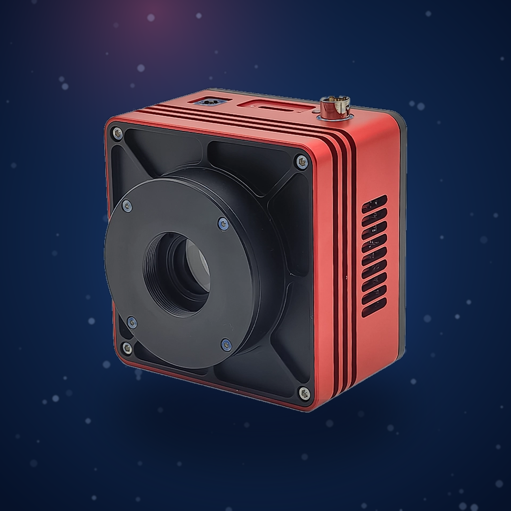
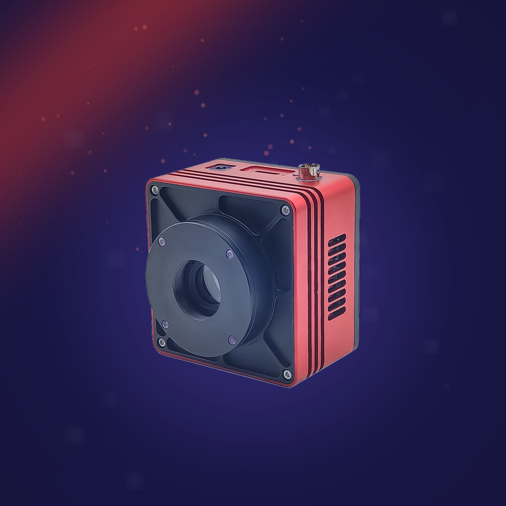
- Imaging Camera: Attostek UVISI064BU High-Sensitivity Camera
Core Advantages: Extremely high quantum efficiency, ultra-low read noise and dark current, ensuring high signal-to-noise ratio images even under low-light conditions, making it critical for detecting weak magneto-optical Kerr signals. - Light Source: Smile Shark Polaris Series White LED Flashlight
Features: Incoherent light source, effectively avoids laser speckle interference, uniform illumination spot, low cost. - Core Optical Components: Objective lens, polarizer, half-wave plate, quarter-wave plate, sample stage, etc.
- Sample: Magnetic thin film with weak perpendicular magnetic anisotropy.
IV. Experimental Procedure
- Optical Path Alignment and Light Source Switching: First, use a laser for optical path alignment to ensure normal incidence of the light path. Then turn off the laser and switch to the white LED flashlight as the illumination source.
- Polarization System Extinction Adjustment: In the absence of a sample or in areas of the sample with no magnetic signal, finely adjust the azimuth angles of the half-wave plate and quarter-wave plate to achieve the maximum extinction state (darkest background), at which point the system is most sensitive to polarization rotation.
- Magnetic Domain Imaging: Move the sample into the optical path. Slightly rotate the half-wave plate (deviating from the complete extinction position by a small angle) to introduce a bias point, linearly converting the polarization plane rotation caused by magnetic domains into light intensity modulation, thereby observing bright-dark contrast magnetic domain patterns in the camera’s field of view.
- Image Acquisition and Processing: Select a uniformly illuminated area as the region of interest (ROI). Use camera software or post-processing software to appropriately adjust image contrast and gamma values to optimize the visual display of the magnetic domain structure and highlight features.
V. Experimental Results and Analysis
Result Analysis:
- Imaging Quality Successfully Verified: The experiment successfully obtained clear labyrinthine magnetic domain images. The domain patterns are continuous and meandering, with obvious bright-dark contrast, and typical domain widths are on the order of 1–3 μm. This directly demonstrates the effectiveness of the combined “white LED + Attostek UVISI064BU High-Sensitivity Camera” solution.
- Analysis of the High-Sensitivity Camera’s Role: The spectrum of the white LED is broad, and the light intensity per unit wavelength is far lower than that of a monochromatic laser, while the magneto-optical Kerr signal itself is extremely weak. The ability to obtain clear images under these conditions is primarily attributed to the exceptional sensitivity and low-noise characteristics of the UVISI064BU camera. Its high quantum efficiency ensures efficient photon capture, and its low read noise ensures that the signal is not淹没 by electronic noise, thus outputting high signal-to-noise ratio raw images even under low-light conditions, providing a high-quality foundation for subsequent image processing.
- Advantages of the White Light Source: The overall image is uniform, with no obvious interference fringes or speckle noise, thanks to the incoherent nature of the white LED. This simplifies image interpretation and avoids interference of coherent noise with the real magnetic domain structure, making it particularly suitable for high-fidelity imaging of static domain structures.
- Physical Interpretation: The observed labyrinth domains are a typical equilibrium structure resulting from the competition between perpendicular magnetic anisotropy and demagnetizing energy in the thin film. The domain width, undulation degree, and other features are directly related to intrinsic material parameters such as the magnetic anisotropy constant, exchange stiffness, and film thickness. This clear static image lays the foundation for further quantitative analysis (such as calculating domain wall energy density) or as a reference initial state for dynamic studies.
VI. Conclusion
This experiment successfully built and validated a wide-field magneto-optical Kerr microscopy imaging system based on white LED illumination and the Attostek UVISI064BU High-Sensitivity Scientific Camera. The system successfully observed clear labyrinth magnetic domain structures in a magnetic thin film with weak perpendicular anisotropy.
The experiment demonstrates that:
- The ultra-high sensitivity and low-noise performance of the Attostek UVISI064BU camera are the decisive factors in overcoming the relatively low light intensity of the white light source and successfully extracting the weak magneto-optical signals.
- The use of a white incoherent light source effectively avoids coherent noise, yielding uniform and clear magnetic domain images while significantly reducing system complexity and cost.