I. NIR-II Sub-Window Imaging
Based on differences in absorption characteristics within the NIR-II (900–1700 nm) band, researchers have further subdivided it into sub‑windows including NIR‑IIa (1300–1400 nm), NIR‑IIx (1400–1500 nm), NIR‑IIb (1500–1700 nm), and NIR‑IIc (1700–1880 nm). Among these, the NIR‑IIx band, which lies near the strong water absorption peak, has been considered to attenuate signals and has therefore seen relatively few in vivo imaging studies.
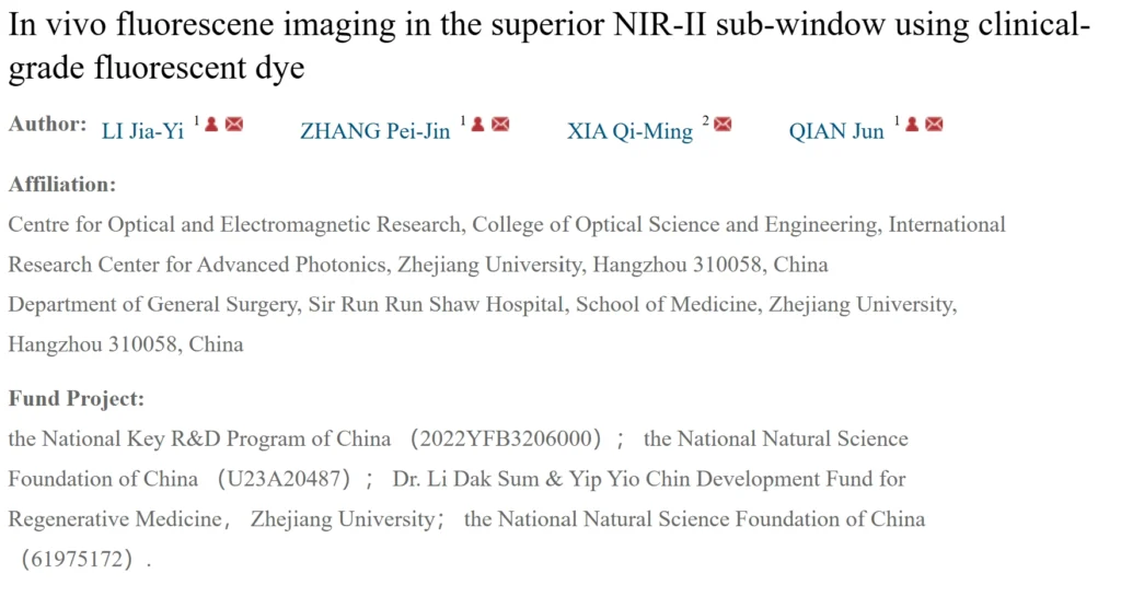
A research team from Zhejiang University systematically conducted in vivo imaging studies in this band using a deep‑cooled SWIR camera. The study suggests that when the fluorescence signal is sufficiently strong, the water absorption characteristics of the NIR‑IIx window can shift from being an “enemy of signal attenuation” to a “friend of background suppression,” enabling high‑contrast imaging. The related findings were published in the Journal of Infrared and Millimeter Waves, 2025, Vol. 44, No. 5.
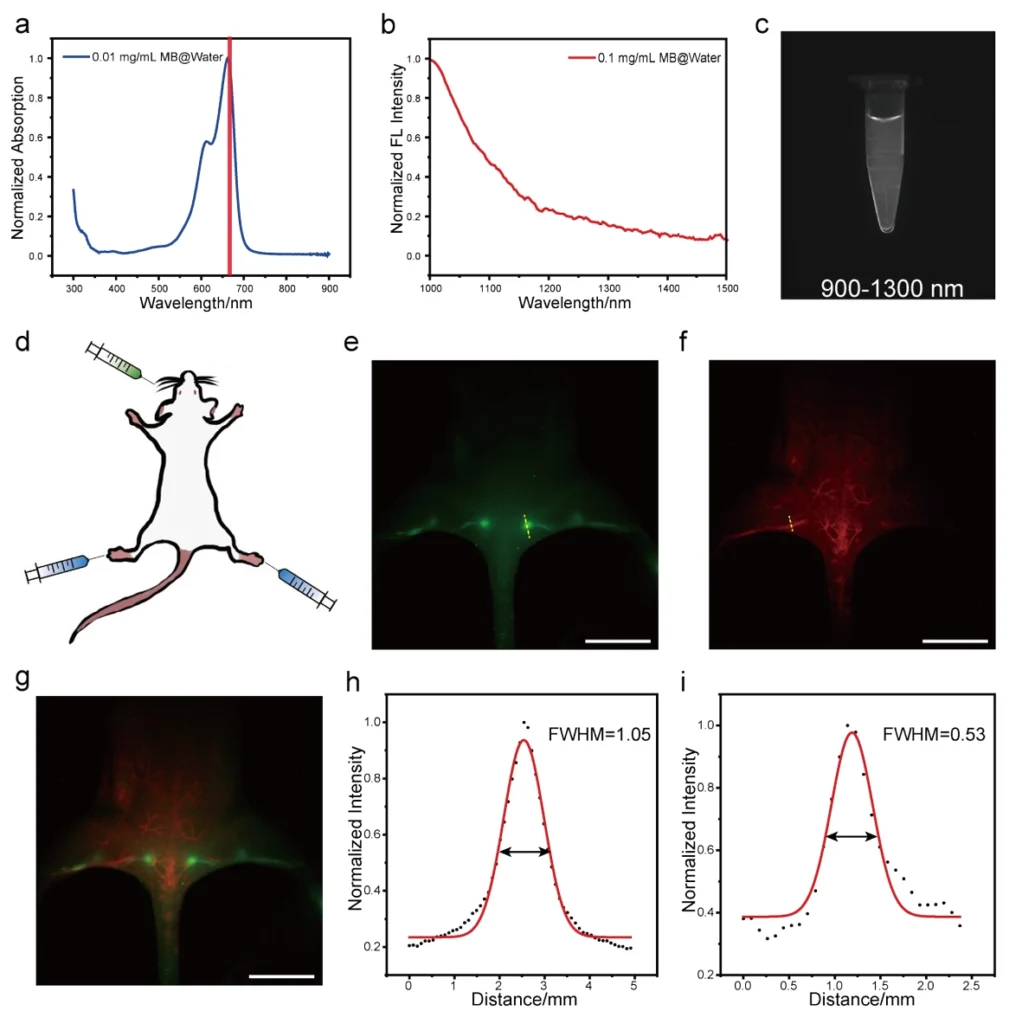
The study systematically compared in vivo vascular imaging quality in mice across four NIR‑II spectral sub‑windows. The experiments first verified theoretical expectations through simulations, then used indocyanine green (ICG) for vascular and intestinal imaging in mice, and employed methylene blue (MB) and ICG for dual‑channel imaging of lymph nodes and blood vessels.
II. Main Research Findings
- The study systematically quantified and demonstrated that the 1400–1500 nm NIR‑IIx window is the optimal sub‑window for ICG vascular imaging, providing a reference for experimental data in this band.
- Standardized protocols for ICG administration parameters, camera acquisition settings, and optical path configuration were established. Dual‑channel imaging enables simultaneous visualization of lymph nodes and blood vessels, providing precise anatomical localization for intraoperative navigation.
- All imaging acquisitions were completed using a domestically produced commercial SWIR camera, providing experimental data support for domestic NIR‑IIx in vivo imaging equipment.
III. Requirements for SWIR Cameras
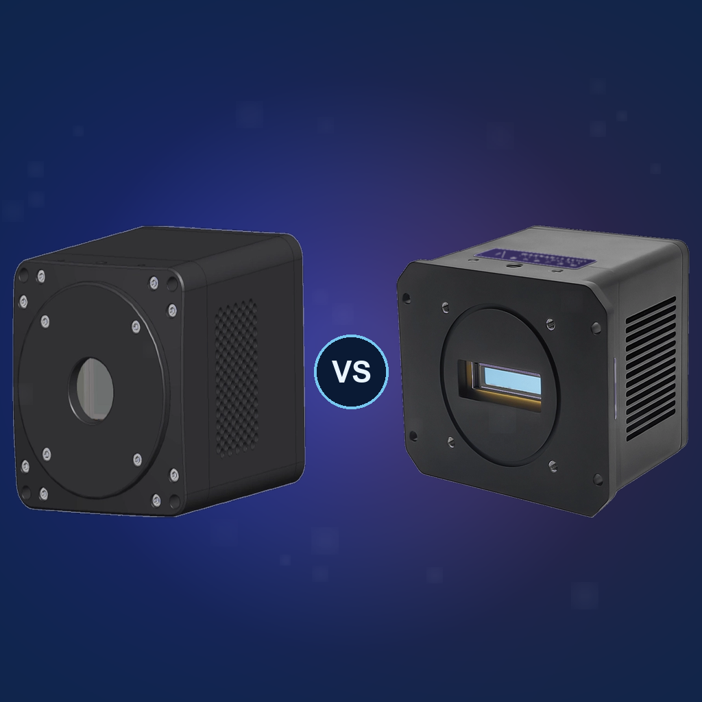
Since the fluorescence signal in the 1400–1500 nm window is extremely weak after attenuation by water absorption, the camera is required to possess extremely high sensitivity and ultra‑low dark current. This study employed a deep‑cooled SWIR camera to suppress dark current, ensuring imaging quality under long‑exposure conditions.
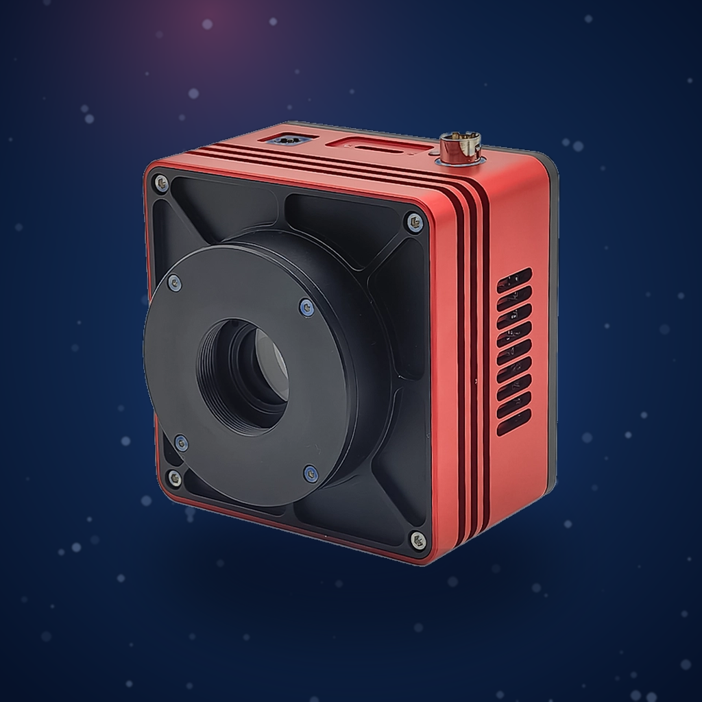
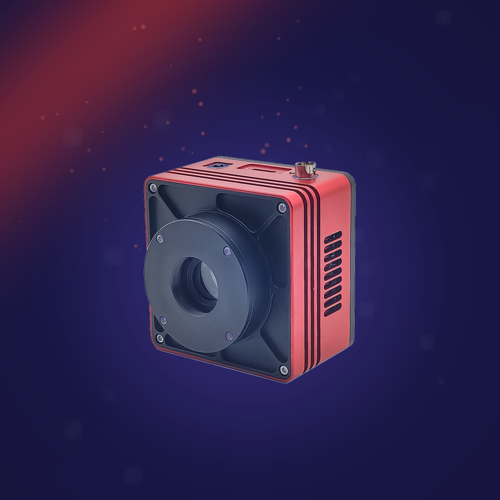
The AttosTek SWIR1503BU‑DCL deep‑cooled SWIR camera covers the 900–1700 nm spectral range and achieves 73% quantum efficiency in the 1400–1500 nm band. Its 15 μm large pixel size effectively captures weak signals. In addition, the ultra‑low dark current enabled by the camera’s –80°C deep cooling better meets the demands of long‑exposure acquisition of weak signals.
IV. Conclusion
With continuous breakthroughs in the performance of deep‑cooled SWIR cameras, in vivo imaging technology based on the NIR‑IIx sub‑window has demonstrated significant potential in clinical scenarios such as tumor surgical navigation, lymph node dissection, and blood supply assessment, thereby accelerating its transition from fundamental research to clinical applications.
Reference: Li J Y, Zhang P J, Xia Q M, Qian J. In vivo fluorescence imaging in the superior NIR‑II sub‑window using clinical‑grade fluorescent dye[J]. Journal of Infrared and Millimeter Waves, 2025, 44(5): 654‑662.
Copyright Notice: This article is intended to share the application of deep‑cooled SWIR cameras in mouse NIR‑II imaging. Some content is adapted from related published research. Copyright belongs to the original authors. Please cite the original source when referencing.