Fluorescence imaging serves as a mainstream non-invasive technique for visualizing dynamic physiological and pathological processes in live mice. Conventional visible-light and first near-infrared window (NIR-I) imaging suffer from prominent drawbacks: severe photon scattering within biological tissues and intense tissue autofluorescence interference, which preclude high-precision imaging of deep tissues. Compared with visible light and NIR-I, the second near-infrared window (NIR-II) features attenuated photon scattering and weaker biological tissue autofluorescence, enabling clear identification of tiny metastatic tumor lesions. This research has greatly advanced investigations into disease pathogenesis and the progress of pharmaceutical development.
I. Mouse NIR-II Imaging
Fluorescence imaging is a mainstream technique for non-invasively observing physiological and pathological dynamic processes in mice. Traditional visible-light and NIR-I imaging have significant limitations: photons scatter severely in biological tissues, and strong tissue autofluorescence interference makes high-precision imaging of deep tissues impossible. Compared with visible light and NIR-I, the NIR-II region reduces photon scattering effects and lowers tissue autofluorescence intensity, enabling clear identification of tumor metastases. This research has greatly advanced the study of disease mechanisms and drug development.
II. Deep-Cooled SWIR Camera
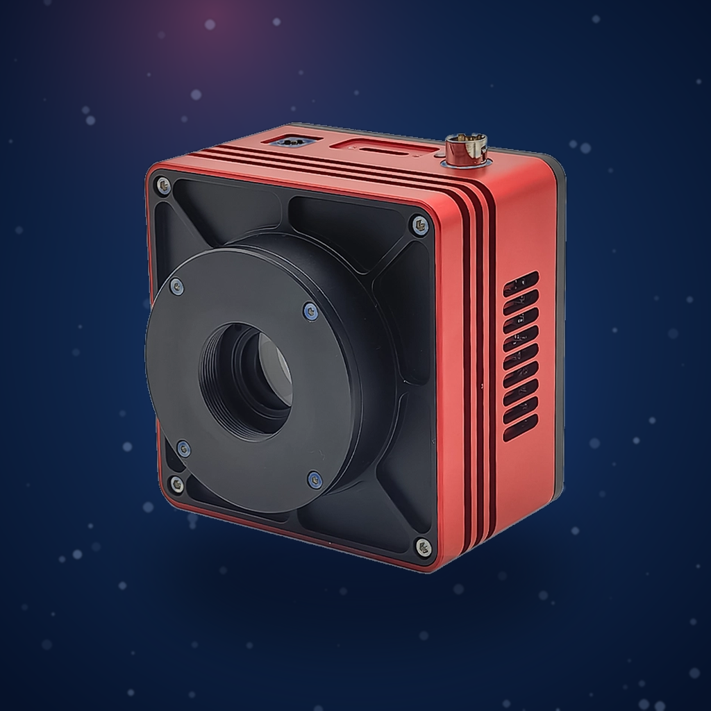
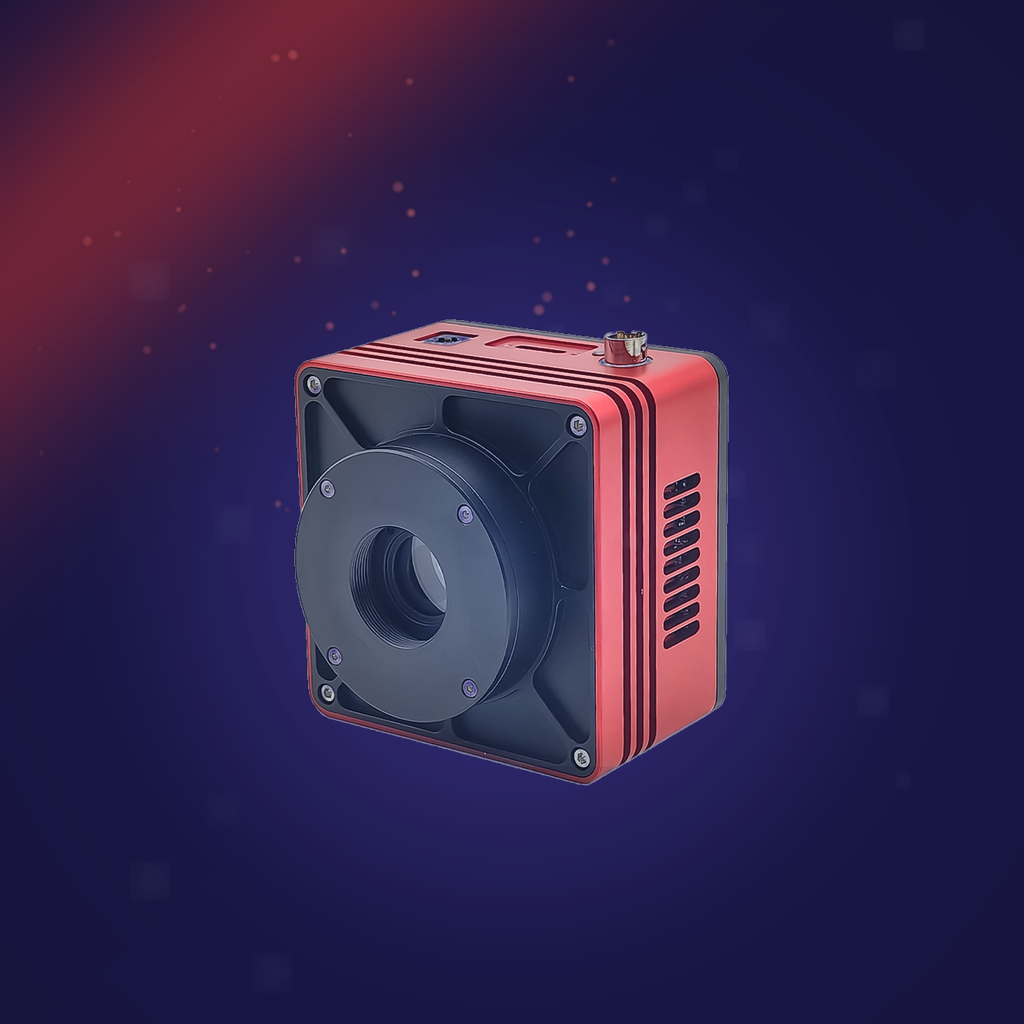
Deep cooling is critical for adapting InGaAs detectors to NIR-II imaging, as it effectively suppresses dark current and enables weak signal capture under long exposures. The AttosTek SWIR1503BU-DC deep-cooled SWIR camera is specifically designed for NIR-II imaging in life sciences, with the following features:
- Equipped with a multi-stage TEC deep-cooling module, the –50°C cooling significantly reduces dark current and background noise, supports long exposure times, and captures extremely weak fluorescence signals from deep mouse tissues.
- Covers the full 900–1700 nm NIR-II band, compatible with mainstream in vivo probes such as ICG, quantum dots, and organic fluorescent dyes, adapting to a variety of imaging experiments.
- The 15 μm large pixel size provides strong photon collection capability, ensuring that even weak signals from deep tissues are clearly captured.
- Global shutter and 300 fps high-speed transmission support dynamic real-time capture and precise quantitative image analysis.
- Standard optical interface is compatible with various scientific lenses and imaging platforms, suitable for custom-built mouse imaging systems and surgical navigation equipment.
III. Typical Application Scenarios
- Tumor lesion imaging: NIR-II fluorescent probes enable precise localization of liver tumors and metastatic lymph nodes. In fluorescence-guided surgery, NIR-II imaging significantly improves tumor boundary delineation and enhances resection precision.
- Vascular structure and blood flow dynamic imaging: By injecting NIR-II fluorescent dyes into mice, high-frame-rate imaging of the entire mouse vascular system can be achieved, with clear resolution of deep organs. In cerebral vascular imaging, it enables complete in vivo vascular imaging of the mouse brain.
- Drug metabolism monitoring: Through fluorescent labeling of drugs, the distribution and metabolic processes of drugs in mice can be directly observed. It also enables in vivo imaging of the mouse intestine and lymphatic system, supporting research on gut microbiota imaging, lymphatic imaging, and pharmacokinetics of macromolecular drugs.
IV. Summary and Technical Specifications
The AttosTek SWIR1503BU-DC deep-cooled SWIR camera, with its InGaAs detector band matching and –50°C deep cooling, is well-suited for a variety of mouse imaging applications, including tumor lesion observation, vascular imaging, and dynamic drug tracking.
SWIR1503BU-DC Camera Core Specifications
| Parameter | Specification |
|---|---|
| Model | SWIR1503BU-DC |
| Sensor Type | InGaAs CMOS |
| Spectral Range | 900 nm – 1700 nm |
| Pixel Size | 15 µm × 15 µm |
| Sensor Format | 3/4″ |
| Frame Rate & Resolution | 300 fps @ 640 × 512 |
| Conversion Gain | 0.96 e⁻/DN (HCG), 5.36 e⁻/DN (MCG), 145 e⁻/DN (LCG) |
| Dynamic Range | 55.8 dB (HCG), 58.1 dB (MCG), 58.3 dB (LCG) |
| Readout Noise | 22.8 e⁻ (HCG), 106 e⁻ (MCG), 2708 e⁻ (LCG) |
| Full Well Capacity | 14 ke⁻ (HCG), 85 ke⁻ (MCG), 2216 ke⁻ (LCG) |
| Maximum SNR | 41.5 dB (HCG), 49.3 dB (MCG), 63.4 dB (LCG) |
| Dark Current | MCG 193.909 e⁻/s/pixel, HCG 630.516 e⁻/s/pixel, LCG 131.561 e⁻/s/pixel |
| Exposure Time Range | 16 µs – 5 s |
| Shutter Mode | Global shutter |
| Data Interface | USB3.0 |
| Data Format | 8-bit / 16-bit |
| Cooling Temperature | –50°C (at 20°C ambient temperature) |
👉 For more technical details, application cases, and specifications of the SWIR1503BU-DC SWIR camera, please visit:
0.33 MP 15 μm Pixels -50°C Cooled 900-1700nm SWIR Camera