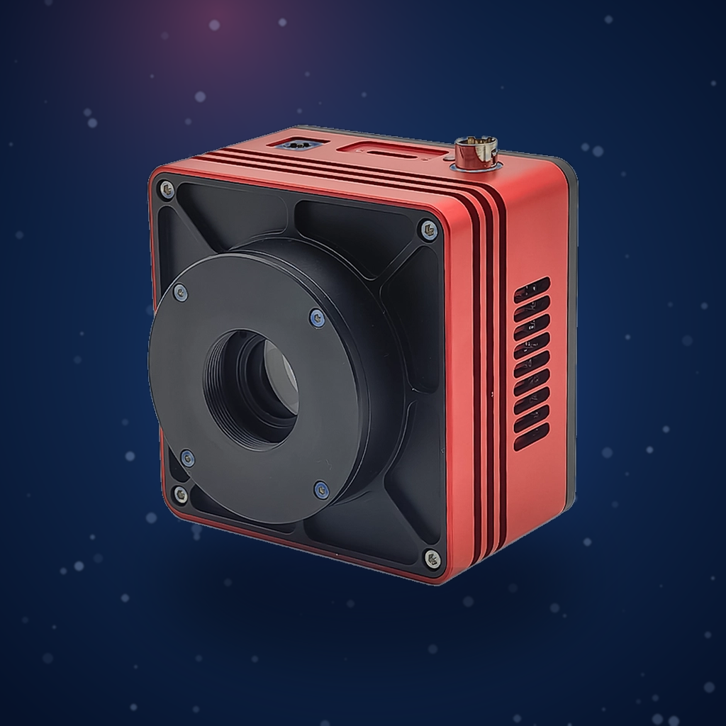
Experiment Name: Single-Molecule Fluorescence Imaging Using the UVISI064BU High-Sensitivity Camera
Experiment Date: November 2023
I. Application Background and Practical Significance
Single-molecule fluorescence imaging is a revolutionary technology in modern life sciences, nanotechnology, and materials science. It enables direct observation of biomolecular behavior, dynamic processes, and nanoscale structures by detecting fluorescence signals at the single-molecule level. Super-resolution microscopy techniques based on single-molecule localization (such as PALM/STORM) have broken the diffraction limit of traditional optical microscopy, pushing spatial resolution to the nanometer scale.
Technical Challenges: Single-molecule fluorescence signals are extremely weak, with each molecule emitting only a limited number of photons (typically a few hundred to a few thousand) per imaging cycle, and the molecules are constantly undergoing photophysical changes (such as blinking and photobleaching). Therefore, achieving high signal-to-noise ratio and high temporal resolution single-molecule detection places extreme demands on the core detector of the imaging system—the scientific-grade camera: extremely high quantum efficiency, ultra-low read noise, and fast, distortion-free electronic readout capability.
Significance of This Experiment:
This experiment aims to evaluate the actual performance of the Attostek UVISI064BU high-sensitivity camera in the critical application scenario of single-molecule fluorescence imaging. By observing the single-molecule fluorescence patterns and dynamic blinking of the classic fluorescent probe Rhodamine 6G, the camera’s core capabilities in single-photon detection sensitivity, temporal resolution, and localization accuracy are directly verified.
Impact on Related Industries and Products:
1. Biomedical Research and Precision Medicine
- Cell Biology and Neuroscience: High-sensitivity single-molecule imaging enables real-time analysis of the nanoscale distribution, aggregation states, and dynamic interactions of proteins on cell membranes (such as receptor clustering and signal transduction), providing molecular-level dynamic images for understanding processes such as synaptic formation, immune responses, and viral infection.
- Pathological Diagnosis and Drug Development: Allows observation of changes in biomarker distribution at the nanoscale, providing new tools for early cancer diagnosis and research on protein aggregation associated with diseases such as Alzheimer’s. At the same time, it enables real-time tracking of drug molecule targeting, binding, and metabolism within cells, accelerating the development and screening of novel targeted drugs.
2. Materials Science and Nanotechnology
- Nanomaterial Characterization: Can be used to study the luminescence uniformity, photostability, and surface modification effects of nanomaterials such as quantum dots, upconversion nanoparticles, and fluorescent polymers, evaluating their performance at the single-particle level.
- Energy and Catalysis: By labeling active sites on catalyst surfaces, single-molecule events during catalytic reactions can be observed, revealing catalytic mechanisms.
II. Experimental Principle
This experiment is based on wide-field fluorescence microscopy and single-molecule detection and localization techniques.
- Single-Molecule Fluorescence Detection: The fluorescent probe (Rhodamine 6G) is diluted to an extremely low concentration (~1 nM) and immobilized on a substrate, so that on average, only one or fewer fluorescent molecules exist within a diffraction-limited spot (approximately 250 nm in diameter). Under excitation by a specific wavelength (561 nm) laser, individual molecules absorb photons and transition to an excited state, subsequently emitting fluorescent photons through radiative decay.
- Point Spread Function and Localization: Light emitted from a single point-like fluorescence source passes through the microscope objective and, due to diffraction effects, forms a two-dimensional Gaussian-distributed spot on the camera plane—this is the Point Spread Function (PSF). The camera’s task is to record this PSF with the highest possible signal-to-noise ratio.
- Fluorescence Blinking: Many fluorescent molecules (including Rhodamine 6G) randomly enter non-fluorescent dark states (such as triplet states) and later return to emitting light—a phenomenon known as “blinking.” Observing single-molecule blinking is an important criterion for distinguishing single molecules from clusters or impurities, and is also a physical mechanism utilized by some super-resolution techniques.
- From Single Molecules to Super-Resolution: By continuously capturing multiple frames of images, recording the random emission and localization processes of a large number of single molecules, and overlaying all these precisely localized molecular positions (with accuracy far below the diffraction limit) onto a single image, a reconstructed image of the sample structure with resolution far exceeding the diffraction limit can be obtained.
III. Task Description
Disperse and immobilize fluorescent probe molecules at low concentration onto a coverslip surface. Under excitation at the corresponding excitation wavelength, a high-sensitivity camera can record the fluorescence patterns of individual fluorescent probes, while also capturing the temporal brightness changes (blinking) of the fluorescence signals from single probes.
IV. Test Equipment and Main Parameters
Key Performance Expectations: High quantum efficiency (especially in the visible spectrum), low read noise (< 1.5 e⁻), and high full-frame readout frame rate.
- Imaging System: Inverted fluorescence microscope equipped with a high-numerical-aperture oil-immersion objective.
- Excitation Light Source: 561 nm continuous-wave semiconductor laser for exciting Rhodamine 6G.
- Fluorescent Probe: Rhodamine 6G, a commonly used single-molecule fluorescence calibration standard with an excitation peak of approximately 525 nm and an emission peak of approximately 550 nm.
- Sample: 1 nM Rhodamine 6G aqueous solution spin-coated onto a clean coverslip surface.
V. Experimental Procedure
- Sample Preparation: Drop 1 nM Rhodamine 6G solution onto a clean coverslip and spin-coat at an appropriate speed to form a sparsely adsorbed monolayer on the surface. Allow to dry for later use.
- System Setup and Alignment: Place the sample on the microscope stage. Install the 561 nm laser excitation module, corresponding dichroic mirror, and emission filter in the optical path. Carefully adjust the optical path to ensure uniform illumination and conjugation with the imaging path.
- Imaging and Data Acquisition:
- Set appropriate exposure time (e.g., 10–50 ms), gain, and readout mode in the camera control software.
- Search for a field of view with sparse fluorescent molecules and clear signals.
- Continuously acquire multiple frames of image sequences to record the appearance, blinking, and photobleaching processes of single-molecule fluorescence spots.
VI. Experimental Results and Analysis
Experimental Results: (Rhodamine 6G, single-molecule fluorescence imaging)
Results Analysis:
- Successful Verification of Single-Molecule Detection: In single-frame images, multiple isolated, bright, approximately circularly symmetric diffraction-limited spots can be observed. These spots are separated from each other, consistent with the characteristics of sparse single-molecule distribution. The spots exhibit high signal-to-noise ratio (SNR) with a uniform and clean background, directly demonstrating that the UVISI064BU camera possesses exceptional sensitivity and extremely low noise levels, enabling effective capture of the limited photons emitted by individual fluorescent molecules within a short time.
- Observation and Analysis of Single-Molecule Characteristics:
- “On-Off” Blinking Phenomenon: Time-trajectory analysis of individual fluorescent spots reveals typical random “on-off” switching in fluorescence intensity—this is blinking. This is a hallmark behavior of single molecules rather than fluorescent clusters or autofluorescence. The camera’s fast frame rate ensures accurate capture of these rapid photophysical dynamics.
VII. Conclusion
This experiment successfully achieved clear imaging and dynamic observation of single Rhodamine 6G molecules using the Attostek UVISI064BU high-sensitivity camera on a wide-field fluorescence microscope. The experiment not only observed isolated diffraction-limited spots consistent with single-molecule characteristics but also captured their signature fluorescence blinking and single-step photobleaching processes.
Conclusions are as follows:
- Performance Meets Requirements: The Attostek UVISI064BU camera’s performance in single-photon detection sensitivity, temporal response, and low-noise control fully meets—and even exceeds—the stringent requirements of single-molecule fluorescence imaging and localization-based super-resolution microscopy.
- Core Value Demonstrated: The camera’s high performance is the fundamental guarantee for successfully achieving single-molecule detection and holds promise for further enabling nanoscale super-resolution imaging. Its reliability as a core component has been verified.