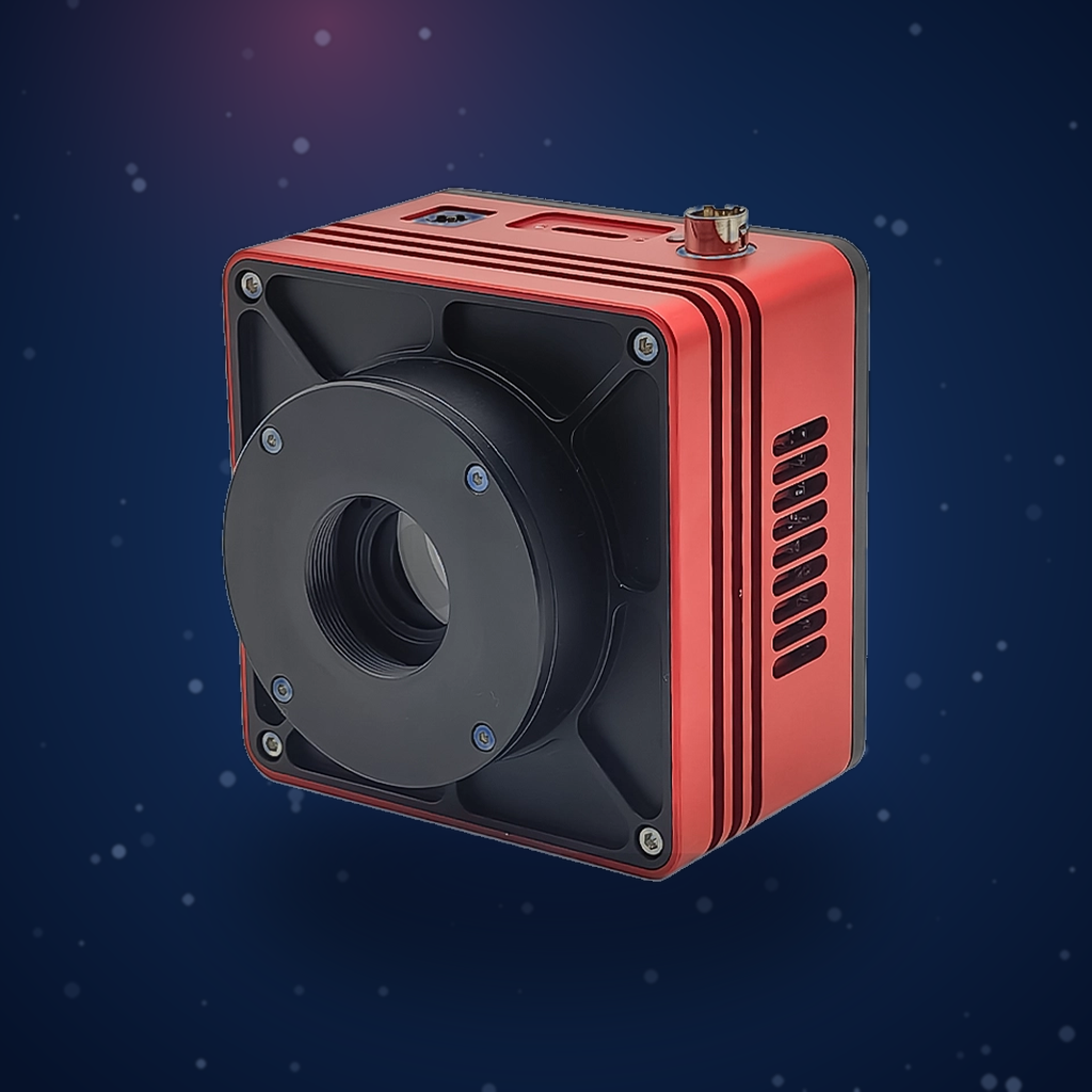
Experiment Title: Imaging Metastable Helium Atoms in a Magneto-Optical Trap Using the Attostek SWIR1503BU Camera
Institution: Institute of Advanced Light Source Facilities, Shenzhen
Experiment Date: April 2026
Ⅰ. Application Background
Metastable helium atoms have an extremely long lifetime and very high energy, making them widely used in fields such as precision measurement and quantum science. Among them, optical pumping magnetometers made from metastable helium atoms are widely applied in geophysical exploration, ocean detection, and military anti-submarine operations. Magneto-optical traps (MOT) can capture helium atoms, and by capturing fluorescence with a camera, it is possible to observe the position and state of helium atoms.
Therefore, it is required that the camera have a high SNR, high frame rate, and high quantum efficiency. The SWIR1503BU camera from Attosecond Technology has a maximum SNR of 62.98 dB at high conversion gain. At 8-bit data format, the frame rate is 724 fps, and its quantum efficiency is about 70% at the wavelength of 1083 nm.
Ⅱ. Task Description
Capturing images of metastable helium atoms being trapped in a MOT using SWIR camera.
In MOT, atoms are confined at the center while continuously absorbing and emitting photons. In this process, atoms scatter faint fluorescence in all directions. By capturing this fluorescence with a camera, the position and state of helium atoms in the MOT can be obtained.
Ⅲ. Testing Equipment and Main Parameters
- Camera:Attostek SWIR1503BU SWIR Camera
- Laser:1083 nm narrow linewidth infrared laser
Ⅳ. Experimental Process
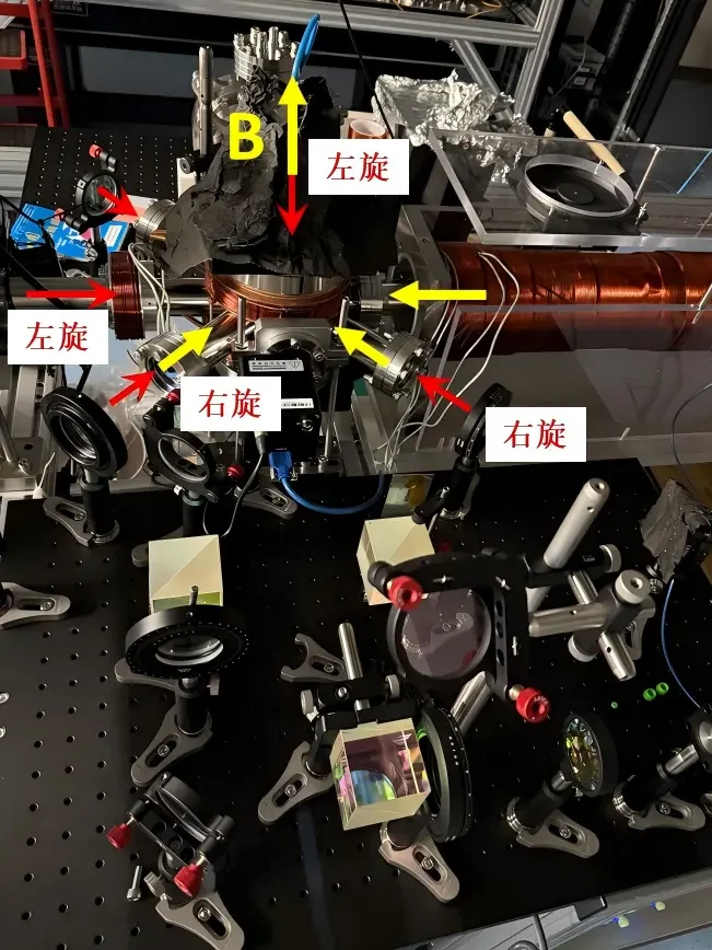
- Use an optical phase-locked loop method to lock the frequency of the 1083 nm laser and split the beam into six paths.
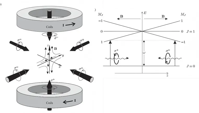
- Direct the six beams into the six directions of the MOT vacuum chamber, adjusting the half-wave plate and quarter-wave plate so that the laser is circularly polarised.
- Switch on the magnetic field, adjust the camera position, and locate the intersection region of the six laser beams.
- Use the camera to observe the captured image of metastable helium atoms. Select the appropriate ROI region and optimise the intensity of the atomic cloud.
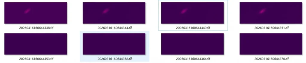
- Using the optical coupling external trigger function of the camera to capture the decay process of atoms in the trap. Although the camera can set the exposure time down to hundreds of microseconds, the interval between captured photos is not that fast. Currently, the shortest exposure time can only be set to 5 ms, with the actual capture generally around 7 ms.
Ⅴ. Experimental Results and Analysis
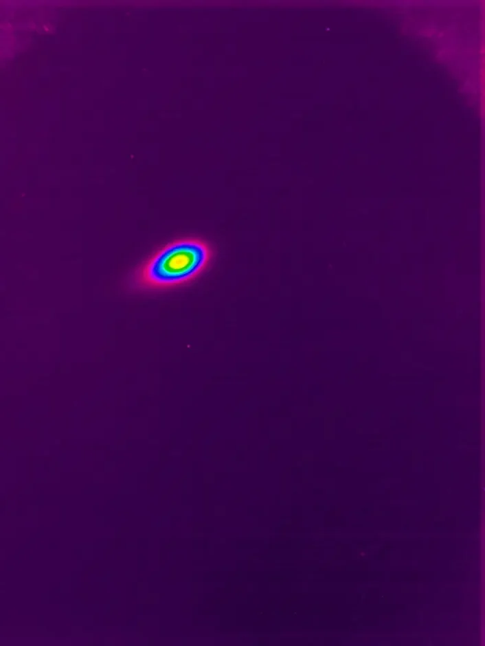
The camera can clearly capture the intensity of 1083 nm fluorescence, observing a bright and stable elliptical light spot with a clear and clean background, demonstrating that metastable helium atoms are confined at the center of the MOT. With rapid, continuous multiple exposures, the fluorescence intensity in the images gradually diminishes until it disappears, showing the decay process of helium atoms in the MOT. Imaging confirms that the SWIR1503BU camera performs stably, with a high SNR and fast readout speed, effectively meeting the requirements of this application scenario.